ABSTRACT
The knee adduction moment (KAM) is commonly used as a surrogate measure of medial compartment loading in individuals with medial knee osteoarthritis. This study investigated the effects of four gait modification strategies—toe-in, toe-out, trunk lean, and knee thrust—on KAM peak and impulse using a within-subject design. Fourteen healthy adults performed normal walking and each modified gait condition. All gait modifications significantly reduced KAM peak compared to normal gait (p<0.05). However, a significant reduction in KAM impulse was observed only during the toe-out gait (p<0.01). Multiple regression analysis revealed that the moment arm accounted for 89–92% of the variance in KAM peak, while the combined effects of moment arm and stance time explained 87% of the variance in KAM impulse (p<0.001). The decrease in impulse during toe-out gait was primarily driven by a lower KAM peak without a significant increase in stance time. In contrast, for the other gait modifications, reductions in KAM peak were counterbalanced by prolonged stance time, resulting in no overall reduction in impulse. These findings suggest that both KAM peak and impulse should be considered when selecting gait modification strategies, with toe-out gait appearing to offer the most favorable biomechanical response in healthy adults.
-
KEYWORDS: Toe-in gait, Toe-out gait, Trunk-lean gait, Knee-thrust gait, Knee adduction moment, Impulse
-
KEYWORDS: 외반보행, 내반보행, 몸통기울임보행, 무릎찌르기보행, 무릎내전모멘트, 충격량
NOMENCLATURE
OA = Osteoarthritis
KAM = Knee Adduction Moment
MA = Moment Arm
GRF = Ground Reaction Force
FPA = Foot Progression Angle
1. Introduction
Osteoarthritis (OA) of the knee is a degenerative condition characterized by the gradual wear of the articular cartilage, leading to pain and joint deformity [
1]. Approximately 25% of individuals aged 55 and older experience knee pain, and among these, about 10% are diagnosed with disabling knee osteoarthritis [
2]. Although total joint replacement is a viable surgical treatment option, prosthetic joints have an average lifespan of approximately 13-14 years [
3]. Therefore, the prevention of knee osteoarthritis and the slowing of its progression have become critical challenges.
Knee OA primarily affects the anterior compartment (patellofemoral joint) and the medial and lateral compartments of the tibiofemoral joint, with the medial compartment being the most frequently affected [
4]. Accordingly, reducing the load on the medial knee to delay cartilage and bone degeneration has been proposed as a preventive and therapeutic strategy for medial knee OA. However, in vivo measurement of medial knee loading is only feasible in patients implanted with prostheses equipped with intra-articular sensors [
5,
6], making this method impractical for widespread clinical application.
As an in vivo–accessible surrogate for medial loading, the knee adduction moment (KAM) has been used [
7-
9]. As justification for its use, Miyazaki (2002) reported that the risk of radiographic progression of medial knee OA (as reflected by joint space narrowing) increased 6.46-fold over a six-year period with a 1% [BW∙H] increase in KAM peak [
7]. Therefore, KAM is closely associated with medial knee loading and the progression of medial knee OA. While earlier studies primarily focused on KAM peaks, more recent research suggest that the time-integral of KAM, known as KAM impulse, may provide a more accurate estimate of OA progression and severity [
9-
12], and that KAM impulse correlates more strongly with medial knee loading than KAM peak [
6]. KAM peaks and impulse represent, the instantaneous maximum medial load and the cumulative load over a single gait cycle, respectively; thus, it is advisable to consider both metrics [
9]. Furthermore, as the step length may vary across individuals and gait strategies, it is reasonable to assess the cumulative load over an identical walking distance, that is, normalized to distance.
Previous studies [
13-
16] have reviewed the literature on gait modification strategies, among which toe-in, toe-out, trunk-lean, and knee-thrust gait patterns have shown particularly effective reductions in KAM. Trunk-lean gait [
17-
22] and knee-thrust gait [
18,
19,
23,
24] have demonstrated significant decreases in KAM or KAM impulse. Likewise, toe-in and toe-out gait modifications have also been reported to effectively reduce KAM [
17,
19,
24-
32].
Determining which gait modification yields the greatest reduction in medial knee load is a critically important question from both academic and clinical perspectives. However, direct comparisons among gait modifications have been limited due to variations in subject characteristics across studies. To address these limitations, it is desirable that the same participants perform multiple gait modifications, allowing for within-subject comparisons of knee loading parameters.
To minimize potential confounding factors, healthy participants were recruited to reduce the influence of uncontrollable variability. In patients with knee osteoarthritis (OA), variables such as lower-limb malalignment and structural deformities can markedly increase inter-individual variability, making it difficult to isolate the true effect of gait modification [
20]. In addition, previous systematic reviews [
13-
16] have synthesized findings from heterogeneous samples, thereby limiting the ability to identify the pure biomechanical effect of gait modification itself. To address these issues, each participant in the present study performed all four gait modifications, which served to minimize inter-individual variability and provide a clearer understanding of their direct biomechanical effects. Nonetheless, as this study involved only healthy adults, compensatory movement patterns and joint loading characteristics specific to OA populations were not captured.
Furthermore, a key methodological inconsistency in prior studies has involved the mixture of unilateral and bilateral gait modification approaches, further complicated direct comparisons. Bilateral gait adjustments have been shown to enhance adherence to instructed modifications [
19,
29], whereas unilateral modifications place an increased burden on the contralateral limb [
33-
35] and may therefore contribute to the progression toward bilateral OA [
34]. Accordingly, the present study adopted a bilateral modification approach to ensure consistency across all gait conditions. In addition, controlling walking speed to be identical across gait strategies may induce unnatural gait patterns, thereby introducing biases that deviate from real-world walking conditions.
Therefore, the present study was designed as an exploratory investigation to compare the changes in the peak values and impulses of the KAM across four gait modification strategies using a within-subject design in healthy adults, while maintaining naturalistic bilateral gait modifications within a laboratory setting, and identifying the common biomechanical mechanisms contributing to these changes.
2. Methods
2.1 Experiments
This study was approved by the institutional ethics committee prior to the experimental procedures (IRB #: 7001355-202011-HR40).
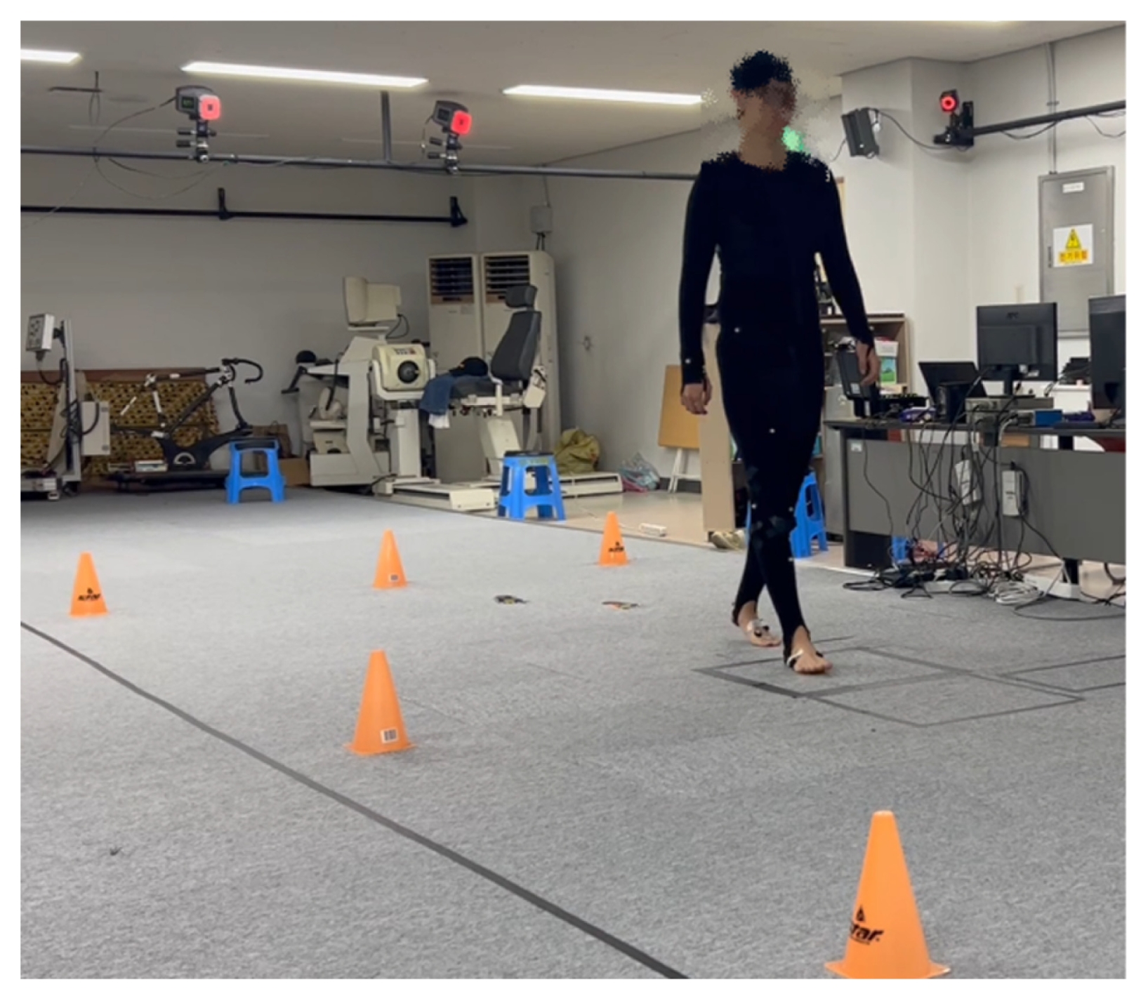
Fig. 1 illustrates the experimental setup utilized in this study. Gait data were collected in a motion analysis laboratory using a 6.5 m walkway equipped with nine motion capture cameras (Eagle, Motion Analysis Corp., USA) and two force plates (AMTI Inc., CA). Participants wore motion capture suits (MS-FS, CS, and WB; 3X3 Designs, Canada) and the Helen Hayes marker set was employed. Fourteen healthy adult men and women were recruited (
Table 1). The inclusion criteria specified healthy adults in their twenties who were able to walk independently without assistive devices, whereas the exclusion criteria specified individuals who reported significant pain or discomfort during the modified gait.
Participants performed at least ten trials per gait strategy, with a 10-minute rest interval between trials. Data collection was performed after a familiarization period exceeding 10-min for each gait strategy. Walking speed was not controlled, and modifications were instructed to be bilateral, as self-selected walking speed and bilateral gait adjustments have been shown to enhance adherence to the instructed modifications [
19,
29].
Instructions for each gait modification were as follows, and all instructions were delivered verbally. Toe-in and toe-out: Bilaterally modify the foot progression angle (FPA) by -10° (toe-in) and +10° (toe-out), respectively, relative to each participant’s natural FPA. Trunk-lean: During the stance phase, lean the trunk 10° toward the stance limb, implementing the same strategy bilaterally. Knee-thrust: Try to rub the medial sides of the knees together while walking [
24], applying the strategy bilaterally. Proper execution of all gait conditions was visually assessed by two independent researchers.
Participants performed the gait conditions in a fixed order (normal, toe-out, toe-in, trunk-lean, and knee-thrust). This sequence was chosen to minimize potential fatigue effects by placing gait conditions that required greater postural adjustment later in the protocol. In addition, to reduce possible carry-over effects between gait strategies, a 10-minute rest and a consecutive 10-minute practice session were provided before the measurements for each gait strategy. For each gait strategy, participants performed ten consecutive trials after the practice session.
2.2 Analysis
Gait data were extracted during the stance phase of the left foot. Among the ten recorded trials, seven with minimal marker loss were selected for subsequent analysis.
Repeated-measures ANOVA and post-hoc tests (LSD) were conducted to determine whether the feature variables in each gait modification significantly differed from those in normal gait. Features included spatiotemporal and kinetic variables related to KAM peaks and impulse.
Spatiotemporal features included walking speed, step width, step length, stance time, and foot progression angle. As for the primary kinetic feature, external KAM was calculated via inverse dynamics analysis using CORTEX software (Motion Analysis, CA). The outcome variables included the two KAM peaks (early and late) and KAM impulse. Impulse was calculated as the time integral of KAM over the stance phase when KAM exceeded zero. Impulse per unit distance was also included, calculated by normalizing impulse by step length. This variable was included to enable more appropriate comparison of cumulative load, as daily cumulative load can be conceptualized as the product of cumulative load per unit distance and walking distance [
36]. Secondary outcomes included ground reaction force (GRF) and moment arm (MA), whose product can approximate KAM [
37]. The moment arm (MA) was calculated as the perpendicular distance between the knee joint center (KJC) and the GRF vector in the coronal plane of the knee joint, following transformation of the coordinates and GRF components into the knee joint’s local coordinate system.
To elucidate the mechanisms underlying the reductions in the three outcome measures (KAM peaks, impulse, and impulse per unit distance), stepwise multiple regression analyses were performed. The entry and removal criteria for independent variables in all models were set at p < 0.05 and p > 0.10, respectively, based on the F-ratio probability.
Independent variables in the regression model for KAM peaks included MA and GRF at the time of each peak. Gait speed was also included in the model, as a reduction in walking speed associated with gait modifications may reduce KAM peaks [
38].
Independent variables in the model for KAM impulse included mean MA, mean GRF, and stance time. Mean MA and mean GRF were defined as the time-averaged values of MA and GRF, respectively, over the period during which KAM exceeded zero. Stance time was included in the model as it defines the interval over which KAM is integrated. Similarly, predictors for impulse per unit distance included mean MA, mean GRF, stance time, and step length.
All statistical analyses were performed using SPSS version 29 (IBM, NY). As this study was designed as an exploratory analysis, we focused on identifying potential patterns and trends in KAM changes across gait modifications without applying strict control conditions. Because this exploratory approach aimed to avoid overlooking potentially meaningful effects, multiple comparison corrections such as the Bonferroni correction were not applied [
39-
43].
3. Result
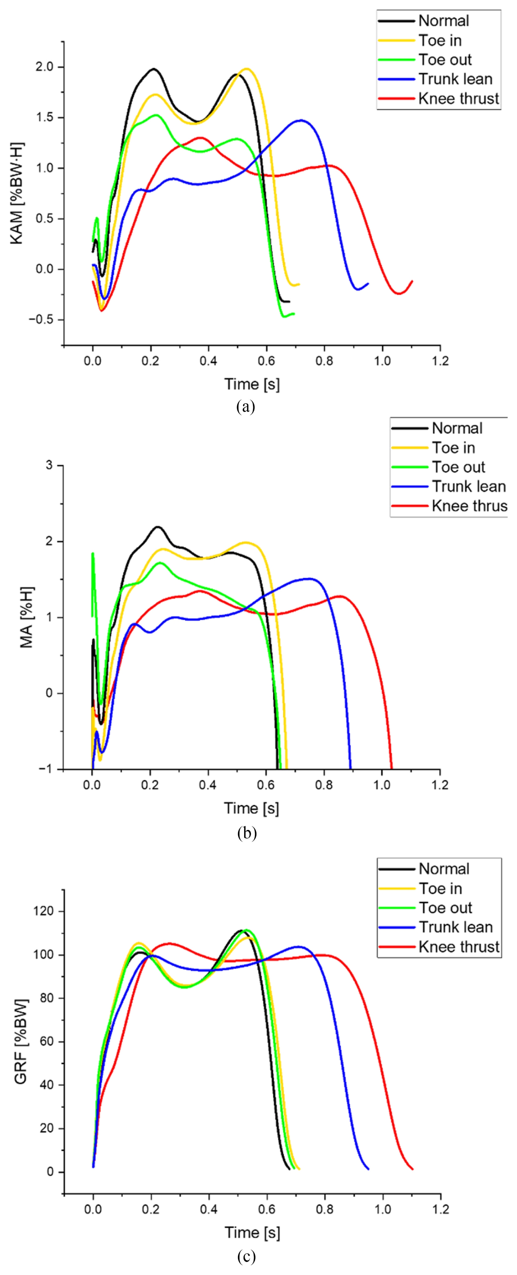
Fig. 2 depicts the KAM waveforms across the stance phase, together with the corresponding MA and GRF waveforms. During gait modifications, the peaks of KAM, GRF, and MA decreased relative to normal gait, whereas an increase in stance time was observed in trunk-lean and knee-thrust gaits (
Figs. 2(a)-
2(c)).
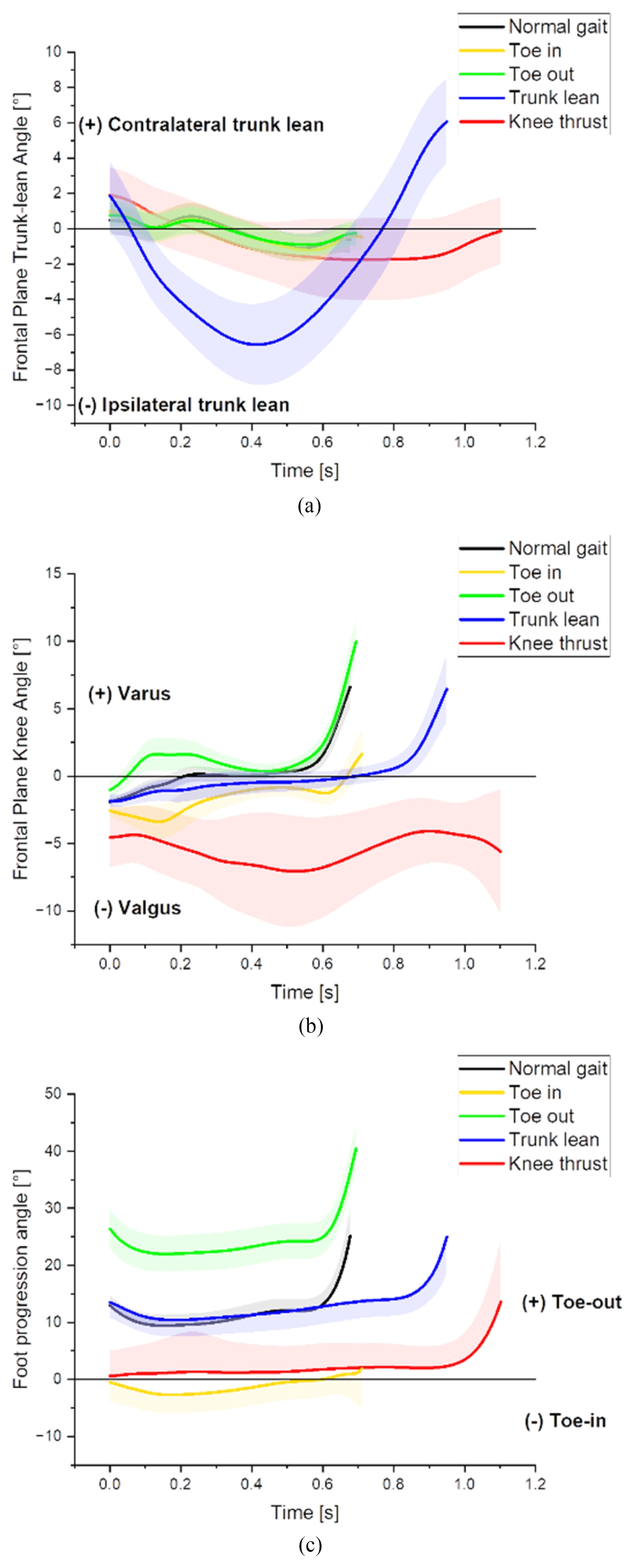
Table 2 and
Fig. 3 present participants’ adherence to the instructed gait modifications, shown in numerical and graphical formats. The foot progression angle changed by approximately +12.5° during toe-out gait and -12° during toe-in gait compared with normal gait (
Table 2). The maximum trunk-lean angle increased by 5.4° ± 2.2° toward the ipsilateral side in trunk-lean gait (
Fig. 3(a)), and the maximum knee valgus angle increased by 5.2° ± 4.1° during the knee-thrust gait (
Fig. 3(b)). These results indicate that the subjects were able to successfully implement each gait modification strategy during the experimental period.
The overall effect sizes of the results were investigated by applying Cohen's guidelines, where a partial eta squared (
np2) of 0.14 or greater indicates a large effect [
44]. The
np2 statistic reflects the proportion of variance in the dependent variable that is explained by a given factor after accounting for error variance. In the present analysis, the
np2 values in
Table 2 mostly exceeded 0.14, indicating large effect sizes. These large
np2 values suggest that the study had sufficient statistical power to detect condition effects even with a relatively small sample size.
Walking speed was reduced, stance time was prolonged, and step width was increased in all gait modifications except toe-out gait (
Table 2). Step length was decreased exclusively in the trunklean gait. Foot progression angle was decreased in toe-in and kneethrust, increased in toe-out, and remained unchanged in trunk-lean.
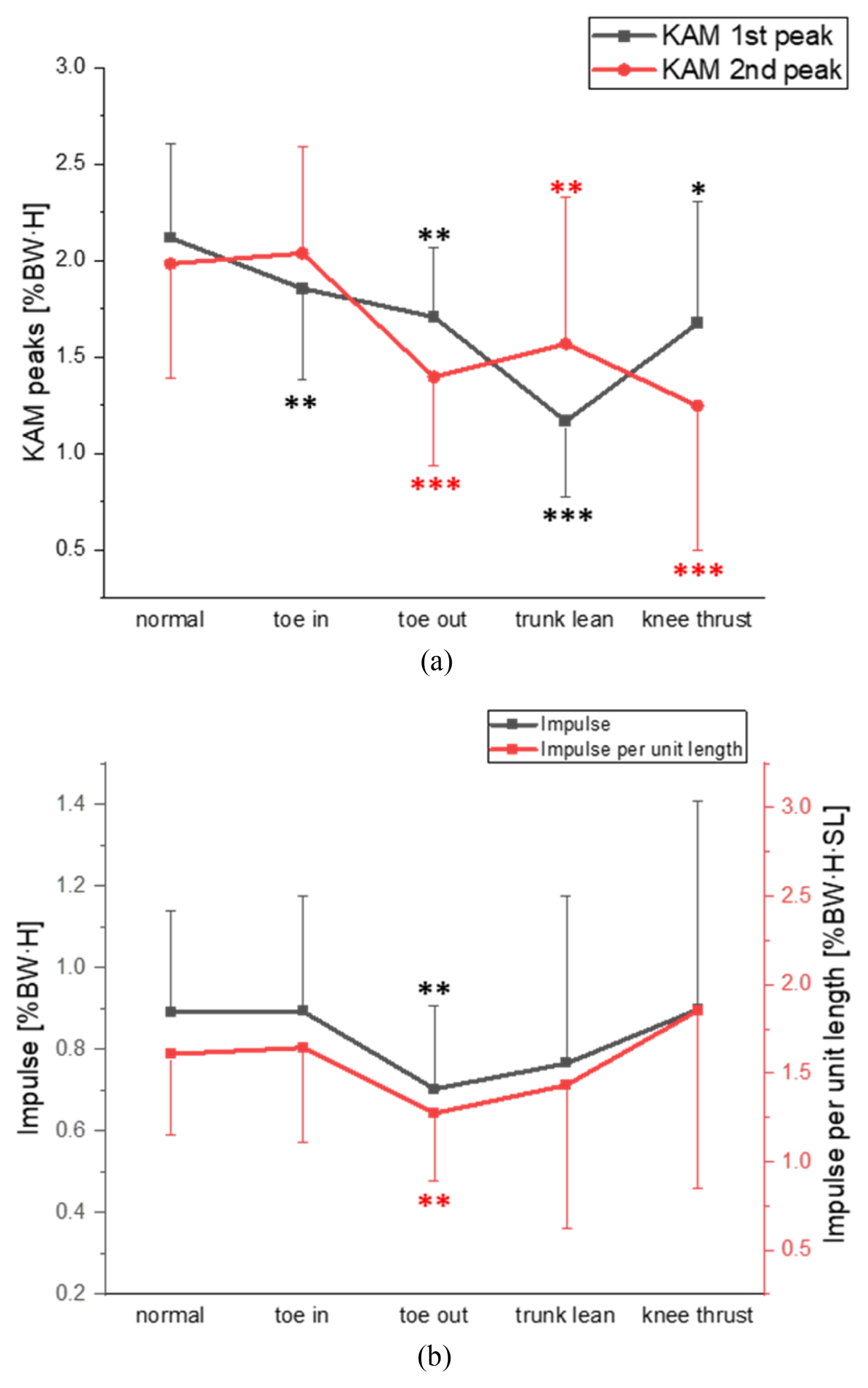
Fig. 4 presents the early and late KAM peaks, the KAM impulse, and the impulse per unit distance. In all gait modification strategies, both KAM peaks were significantly reduced relative to normal gait, with the only exception being the second KAM peak in the toe-in condition (
Fig. 4(a) and
Table 2).
Table 2 compares MA and GRF at peak instants with those of normal gait. At the first peak instant, MA was reduced in all gait modifications except knee-thrust, whereas GRF did not differ significantly. At the second peak instant, MA was reduced in all modifications except toe-in, and GRF was significantly reduced only in trunk-lean and knee-thrust conditions.
In the stepwise multiple regression analysis for KAM peaks, MA alone accounted for 89–92% of the variance, and the addition of GRF to the model increased the explanatory power by only 3– 7% (
Table 3). Gait speed was excluded from the final model during the stepwise process, as the
p-value for the F-statistic exceeded the predefined removal threshold (
p > 0.10).
Among the four gait modifications, both KAM impulse and KAM impulse per unit distance were significantly reduced only during toe-out gait (
Fig. 4(b) and
Table 2).
Table 2 also shows that the mean MA decreased in all gaits except toe-in, the mean GRF decreased in knee-thrust and trunk-lean, but the stance time also increased in all modifications except toe-out.
Table 4 presents the results of the stepwise multiple regression for KAM impulse and KAM impulse per unit distance. Mean MA and stance time together explained 87% of the variance in KAM impulse and 79% of the variance in impulse per unit distance; adding mean GRF and step length to the model yielded only a marginal increase in explanatory power (Δ
R2 = 0.03–0.07).
4. Discussion
4.1 Major Findings
KAM peaks were reduced in most gait modifications (
Fig. 4a). Multiple regression analysis indicated that the decrease in the moment arm (MA) was the strongest factor associated with the reduction in KAM peaks across all gait modifications, with MA alone explaining at least 89% of the variance in KAM peaks (
Table 3).
In contrast, KAM impulse was reduced exclusively during toe-out gait (
Fig. 4b). Given that the combination of mean MA and stance time explained 87% of the variance in impulse (
Table 4), it can be inferred that the prolonged stance time counteracted the reduction in MA in all other gait modifications. KAM impulse per unit distance exhibited the same pattern, likely due to minimal variation in step length across most gait modifications.
KAM impulse has been reported to reflect OA progression and severity more accurately than KAM peaks [
9-
12]. Walter et al. investigated the association between in-vivo medial contact force (MCF) and both KAM peaks and impulse. They found that decreases in impulse closely matched reductions in MCF, whereas decreases in KAM magnitude corresponded to those of MCF at the 2nd peak, but not at the 1st peak [
6]. Therefore, validity of relying solely on the 1st peak KAM as a surrogate marker of medial loading remains questionable. Uhlrich et al. reported that the first peak KAM was greater than the second in 93% of 107 patients with medial OA [
32], which has motivated numerous studies to focus on reducing the first peak KAM. However, targeting the reduction of the first peak KAM, which may not correspond to the decrease in MCF, cannot be regarded as definitely effective.
In the case of normal populations, toe-out gait among the four modifications is considered the most effective strategy for reducing medial knee loading, because only toe-out gait resulted in reductions in both the peak and impulse of KAM. Furthermore, Caldwell et al. (2013) reported that toe-out gait imposed lower cognitive and physical demands compared with other gait modification strategies [
17]. Therefore, toe-out gait is clinically advantageous not only because of its simplicity but also due to its high patient compliance.
The results of this study were compared with those of previous research and are presented in
Table 5. The following sections provide a detailed discussion of each gait modification individually.
There have been several systematic reviews that have synthesized studies examining the effects of gait modification on the KAM and related biomechanical variables [
13-
16]. However, because these reviews compared results from independently conducted studies, inter-individual differences among participant groups make it difficult to accurately evaluate the relative effectiveness of different gait modification strategies, and the underlying mechanisms responsible for KAM reduction have not been directly verified. In contrast, the present study minimized inter-individual variability by having the same participants perform all four gait modification strategies, thereby enabling within-subject comparisons and revealing, through regression analysis, that the reduction in KAM was primarily attributed to a decrease in the MA. Additionally, to reproduce realistic walking conditions in the laboratory, walking speed and step length were not artificially controlled.
In the present study, bilateral gait modification was implemented to enhance participant adherence and ensure the consistent execution of gait patterns. However, this approach may limit direct clinical applicability for patients with unilateral medial knee OA, in whom asymmetrical loading patterns are commonly observed. Individuals with unilateral OA often adopt compensatory strategies, such as increased knee varus and reduced knee flexion on the ipsilateral side, which places an increased burden on the contralateral limb [
33-
35]. Given this elevated loading risk, Jones et al. (2013) suggested that unilateral OA may frequently progress to bilateral involvement [
34]. Thus, bilateral gait modification could potentially offer mechanical or clinical advantages by promoting overall gait symmetry and reducing contralateral overload, although further research is warranted to confirm these effects in asymmetric populations.
4.2.1 Toe-In & Out
Typically, toe-in gait has been reported to reduce the 1st peak of KAM, whereas toe-out gait reduces the 2nd peak [
31]. As a mechanism underlying these KAM reductions, a lateral shift of the center of pressure (COP) — occurring during the early stance phase in toe-in gait and the late stance phase in toe-out gait — which leads to a decrease in MA, has been proposed [
25,
29,
31]. However, mediolateral displacements of COP in the global frontal plane may not always correspond to those in the local frontal plane defined by the knee joint (shank) axes, indicating that the specific mechanisms require more detailed analysis.
The results of the present study for toe-in gait (reduction in 1
st peak and no difference in 2
nd peak) are consistent with those of Shull et al. [
29] and Uhlrich et al. [
31], but differ from the findings of Lynn et al. [
28] and Lindsey et al. [
19] where increases in the second peak KAM were observed. This increase may be explained by excessively large postural adjustments and overly strict control protocols. Specifically, Lynn et al. instructed participants to adopt a toe-in angle of -30°, which may have caused excessive medialization of the foot at the time of the second peak, thereby increasing the GRF moment arm. Lindsey et al. provided real-time feedback of foot progression angle (FPA), trunk angle, and knee angle, requiring participants to maintain these variables within a strict predefined range. They suggested that the unnatural movement pattern induced by such stringent instructions may have contributed to the observed increase in KAM [
19].
All previous studies on toe-out gait have reported a reduction in the second peak of KAM, which is consistent with our findings. However, only our study demonstrated a statistically significant decrease in the first peak of KAM. Uhlrich et al. [
31] also observed a trend toward a reduction in the first peak KAM (p = 0.06) and suggested that increased step width might have contributed to this effect. Our data showed similar increase in step width in toe-out gait, though not significant. Two possible explanations can be proposed. First, most previous studies employed unilateral feedback protocols, and the resulting asymmetry may have induced an unnatural gait pattern that hindered reduction in the first KAM peak. Second, in the bilateral toe-out gait, the external rotation of foot may lead to external rotation of the tibia and this may act to decrease the moment arm of GRF vector around KJC in the frontal plane of tibial axes at the first peak.
In this study, KAM impulse did not differ from that of normal gait during toe-in gait, but was significantly reduced during toe-out gait, which is consistent with previous studies.
4.2.2 Trunk-lean
Gerbrands et al. [
18] suggested that the reduction in KAM observed during a trunk-lean gait might be attributed to a decrease in the MA of the ground reaction force, although this was not verified in their study. In our previous study [
21], we empirically confirmed that the reduction in MA was indeed the main contributor to KAM reduction and further demonstrated that this decrease resulted from the medial displacement of the knee joint center.
In the present study, reductions in KAM peaks were similar to those reported in previous studies, except for those by Caldwell et al. and Lindsey et al. [
17,
19]. This discrepancy may have arisen from the differences in gait speed control. A decrease in gait speed appears to be inherent in trunk-lean gait, as it is a challenging coordinative movement [
18]. Likewise, when the trunk sway angle increases, gait speed naturally decreases to maintain stability and safety. Reduced speed might make the effect of trunk lean pronounced only during the early stance phase, so that the 2
nd peak of KAM may not be reduced. Lindsey et al. [
19] implemented stricter speed control compared to Simic et al. [
22], which likely resulted in an insignificant change in the second KAM peak.
KAM impulse showed a significant reduction in Simic et al. [
22], Caldwell et al. [
17], and Gerbrands et al. [
18], which contrasts with the result of the present study. In Simic et al. [
22] and Caldwell et al. [
17] gait speed was controlled, resulting in a stance time comparable to that of normal gait, which allowed for a reduction in impulse. On the other hand, though gait speed was not controlled in Gerbrands et al. [
18], the reduction in gait speed was small (10.7% compared to 28% in the present study) and impulse decreased. The small change in speed might have been due to the adoption of unilateral trunk-lean strategy.
4.2.3 Knee-thrust
Knee-thrust gait was first developed by Fregly et al. [
23] through model-based simulations. It has been suggested that the medial-thrust gait reduces KAM by decreasing the MA of the GRF vector [
23,
24]. Our preliminary study confirmed the reduction of the MA and its mechanism: the COP shifted considerably farther laterally than the KJC, which resulted in a decreased MA [
45].
The results for peak reduction are consistent with those reported by Fregly [
23] and Schache [
24], but differ from Gerbrands [
18] and Lindsey [
19] for the 2
nd peak. The former instructed bilateral knee-thrusting, whereas the latter employed unilateral one. It appears that the knee thrusting occurs throughout the stance phase in the bilateral condition, whereas in the unilateral condition it is concentrated on the early stance with a diminishes in late stance.
In the present study, both KAM peaks decreased; however, gait speed also declined, resulting in no significant change in KAM impulse. The reductions in impulse observed in previous studies are likely attributable to gait speed control and/or unilateral gait modification. For example, Lindsey et al. [
19] controlled gait speed, allowing the reduction in KAM peaks to translate directly into a reduction in impulse. In contrast, although Gerbrands et al. [
18] did not control gait speed, unilateral strategy resulted in only 15% decrease in gait speed, in contrast to the 41% reduction observed in the present study. Therefore, the smaller increase in stance time may not fully offset the reduction in KAM peaks.
In Schache et al. [
24], a significant reduction in KAM impulse was observed despite the absence of gait speed control and the use of bilateral knee thrust. This may be due to the fact that the study included only a single participant, that is, this individual exhibited a decrease in KAM peaks, but the reduction in gait speed was small, leading to a concomitant decrease in impulse. Similarly, in the present study, one of the 14 participants showed little change in gait speed (normal: 1.00 m/s; knee-thrust: 0.92 m/s) and therefore showed a 59% reduction in KAM impulse compared to normal gait.
First, although the magnitude of KAM has been widely used as an indirect indicator (surrogate measure) of MCF in the knee, KAM alone is insufficient to fully explain MCF [
6]. Previous studies demonstrated that when the knee flexor moment (KFM) was included along with KAM, the resulting regression model for MCF achieved a higher coefficient of determination (
R2) of 0.85– 0.93 [
6,
46]. Furthermore, other biomechanical and pathological factors — including joint alignment, muscle co-contraction, and meniscal degeneration — are also known to influence MCF [
47-
50]. Therefore, acknowledging these additional determinants would be crucial for obtaining a comprehensive understanding of the multifactorial nature of MCF.
Second, despite the finding of large effect sizes (
Table 2), a limitation of this study lies in interpreting these values. The Cohen's guidelines applied for effect size are primarily derived from data distributions in the behavioral sciences [
44]. Given the nature of biomechanical experiments — where experimental control is typically high, and the error variance (SS
Error) is often lower than in behavioral science fields — the
np2 values are likely to be relatively inflated or overestimated. Consequently, the observed effects must be interpreted more conservatively than the conventional thresholds suggest, and recruiting a larger sample of participants in future research is recommended to improve the generalizability of the findings.
Lastly, this study was conducted on healthy individuals; therefore, its findings may not be directly generalizable to clinical populations. Although the toe-out gait was identified as the most effective strategy among the four gait modifications (
Table 2), this result reflects responses in individuals without structural deformities. In patients with anatomical deformities, such as varus malalignment, other gait modifications may yield more favorable biomechanical outcomes. Further studies involving medial knee OA populations are warranted to evaluate the clinical applicability of these findings in therapeutic contexts.
5. Conclusion
During gait modification, reductions in KAM peak were explained by MA by up to 89%, whereas KAM impulse was influenced not only by MA but also by stance time. Among the four gait strategies tested, when implemented following simple verbal instructions without extended training, only the toe-out gait produced significant reductions in both peak and impulse. Therefore, toe-out gait may be considered the most effective strategy for reducing KAM. By directly comparing multiple gait modifications within the same participants and simultaneously evaluating both peak and impulse, this study offers a more comprehensive and systematic comparison of their relative biomechanical effects.
FOOTNOTES
-
ACKNOWLEDGEMENT
This paper was supported by Konkuk University in 2025 and the Basic Science Research Program through the National Research Foundation of Korea (NRF) funded by the Ministry of Education, Korea (No. RS-2021-NR066085).
Fig. 1Experimental setup of the gait analysis laboratory. The 6.5 m walkway was instrumented with nine motion capture cameras and two force plates. Subjects wore a motion capture suit with the Helen Hayes marker set while walking under the predefined experimental conditions
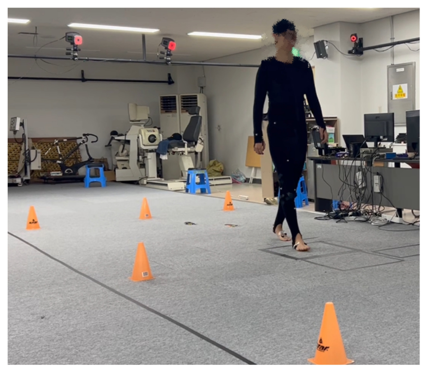
Fig. 2Averaged stance phase waveforms of key kinetic variables (KAM, MA, and GRF) across gait conditions. (a) Knee adduction moment, (b) moment arm of ground reaction force with reference to the knee joint center, and (c) ground reaction force magnitude. The abbreviations of the units used for the Y-axis indicate: BW for body weight, and H for height
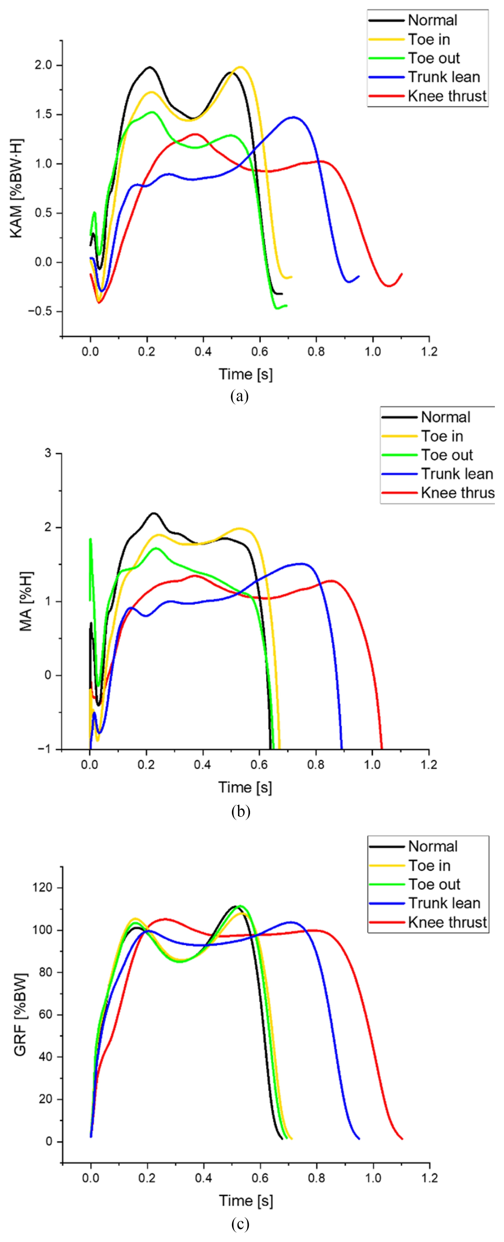
Fig. 3Averaged stance phase waveforms of key kinematic variables across gait conditions. (a) frontal plane trunk-lean angle, (b) frontal plane knee angle, and (c) foot progression angle
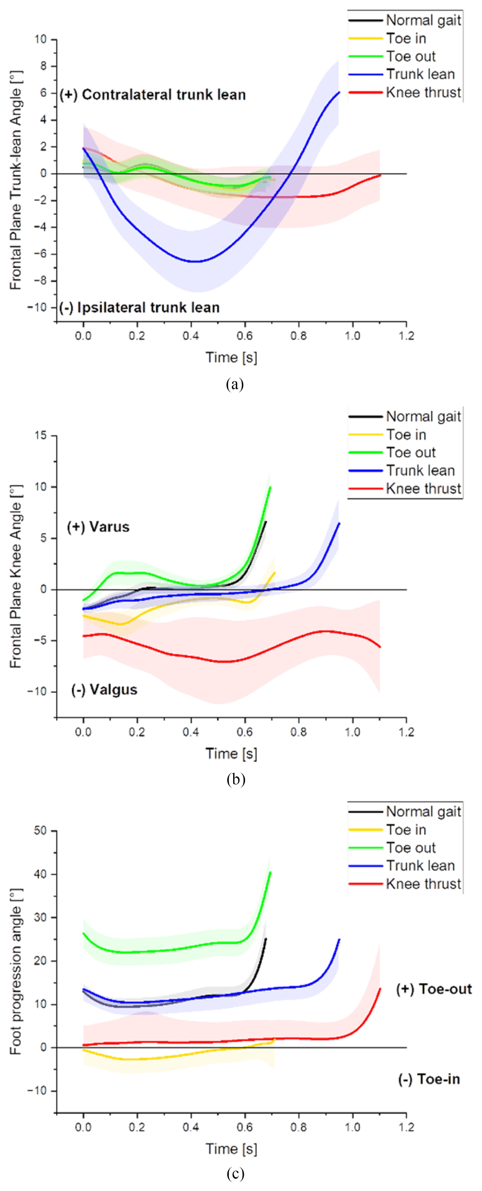
Fig. 4Comparison of outcome variables among different gait types. (a) KAM peaks and (b) KAM impulse and impulse per unit distance. Statistical differences in an outcome variable between a modified gait and the normal gait are shown in each graph: * p < 0.05, ** p < 0.01, *** p < 0.001. The abbreviations of the units used for the Y-axis variable are: BW for body weight, H for height, and SL for step distance
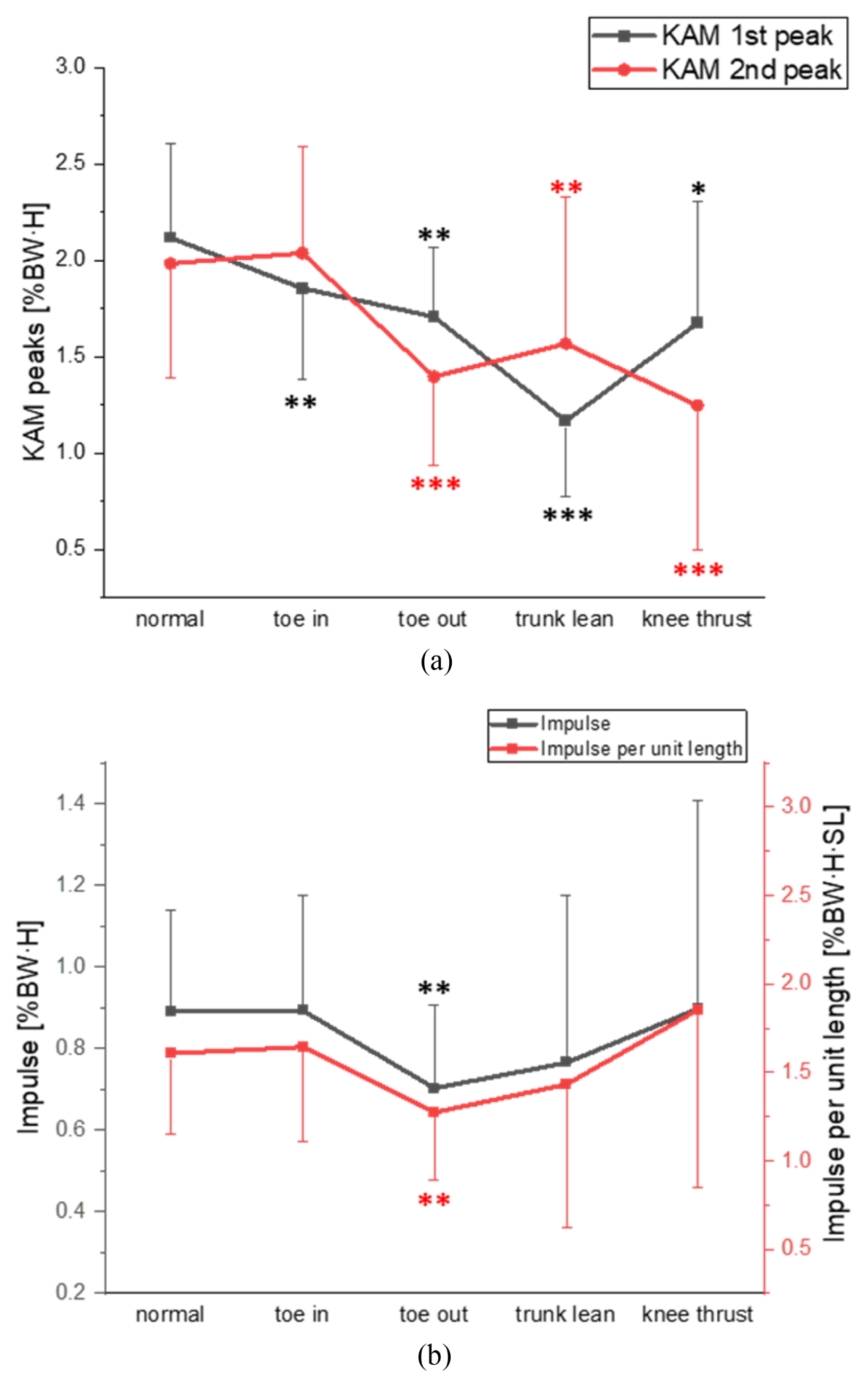
Table 1
Table 1
|
Characteristic |
Mean ± SD (n=14) |
|
Gender |
Male: 7, Female: 7 |
|
Age [yrs] |
22 ± 1.7 |
|
Height [m] |
1.68 ± 0.94 |
|
Body mass [kg] |
67.8 ± 14.3 |
Table 2Comparison of feature variables among different gait types
Table 2
|
Category |
Variable |
Normal |
Toe-in |
Toe-out |
Trunk-lean |
Knee-thrust |
ηp2
|
|
Spatiotemporal features |
Walking speed [m/s] |
0.82 ± 0.70 |
0.77 ± 0.10*
|
0.80 ± 0.90 |
0.59 ± 0.14***
|
0.48 ± 0.17***
|
0.76 |
|
Step width [mm] |
112 ± 28 |
145 ± 41**
|
118 ± 30 |
175 ± 42***
|
212 ± 39***
|
0.68 |
|
Step length [mm] |
556 ± 20 |
547 ± 27 |
556 ± 27 |
537 ± 32*
|
499 ± 101 |
0.19 |
|
Stance time [s] |
0.67 ± 0.04 |
0.71 ± 0.07*
|
0.69 ± 0.05 |
0.94 ± 0.22***
|
1.10 ± 0.28***
|
0.66 |
|
Foot progression angle [°] |
9.5 ± 6 |
−2.5 ± 7***
|
22 ± 7***
|
10 ± 7 |
1 ± 7***
|
0.84 |
|
|
Primary kinetic outcomes |
KAM 1st peak [%BW·H] |
2.12 ± 0.49 |
1.86 ± 0.47**
|
1.71 ± 0.36**
|
1.17 ± 0.39***
|
1.67 ± 0.63*
|
0.48 |
|
KAM 2nd peak [%BW·H] |
1.98 ± 0.60 |
2.04 ± 0.55 |
1.4 ± 0.46***
|
1.57 ± 0.76**
|
1.25 ± 0.75***
|
0.46 |
|
KAM impulse [%BW·H] |
0.89 ± 0.24 |
0.89 ± 0.28 |
0.70 ± 0.20**
|
0.76 ± 0.41 |
0.89 ± 0.50 |
0.11 |
|
KAM impulse per unit length [%BW·H·SL] |
1.60 ± 0.46 |
1.64± 0.53 |
1.27 ± 0.38**
|
1.43 ± 0.8 |
01.85 ± 1.00 |
0.17 |
|
|
Secondary kinetic variables |
MA at 1st peak [%H] |
2.31 ± 0.50 |
2.01 ± 0.50**
|
1.93 ± 0.41**
|
1.38 ± 0.47***
|
1.75 ± 0.81p = 0.05
|
0.34 |
|
GRF at 1st peak [%BW] |
97 ± 4 |
96 ± 7 |
92 ± 9 |
91 ± 7**
|
101 ± 5 |
0.34 |
|
MA at 2nd peak [%H] |
1.94 ± 0.50 |
2.06 ± 0.54 |
1.36 ± 0.40***
|
1.63 ± 0.78*
|
1.36 ± 0.89*
|
0.36 |
|
GRF at 2nd peak [%BW] |
108 ± 7 |
107 ± 6 |
106 ± 8 |
101 ± 5***
|
100 ± 8***
|
0.44 |
|
Mean MA [% H] |
1.48 ± 0.38 |
1.38 ± 0.36 |
1.15 ± 0.28***
|
0.91 ± 0.47***
|
0.95 ± 0.41**
|
0.44 |
|
Mean GRF [% BW] |
79 ± 2 |
78 ± 2 |
78 ± 3 |
72 ± 9**
|
70 ± 11*
|
0.30 |
Table 3Stepwise multiple regression analysis of the KAM peaks
Table 3
|
Dependent variable |
Factors |
Final adjusted R2
|
Δ Adjusted R2
|
VIF |
β (95% CI) |
|
KAM 1st peak [%BW·H] |
MA [%H] |
0.96***
|
0.89 |
1 |
0.92***(0.8–0.9) |
|
GRF [%BW] |
0.07 |
1 |
0.26***(0.2–0.3) |
|
Speed [m/s] |
Excluded |
|
|
|
KAM 2nd peak [%BW·H] |
MA [%H] |
0.95***
|
0.92 |
1.2 |
0.87***(0.8–0.9) |
|
GRF [%BW] |
0.03 |
1.2 |
0.18***(0.1–0.2) |
|
Speed [m/s] |
Excluded |
|
Table 4Stepwise multiple regression analysis of the KAM impulse & KAM impulse per unit distance
Table 4
|
Dependent variable |
Factors |
Final adjusted R2
|
Δ Adjusted R2
|
VIF |
β (95% CI) |
|
KAM impulse [%BW·H] |
Mean MA [%H] |
0.90***
|
0.57 |
1.4 |
0.82***(0.7–0.9) |
|
Stance time [s] |
0.30 |
1.2 |
0.63***(0.5–0.7) |
|
Mean GRF [%BW] |
0.03 |
1.6 |
0.21***(0.1–0.3) |
|
|
KAM impulse per unit distance [%BW·H·SL] |
Mean MA [%H] |
0.89***
|
0.44 |
1.5 |
0.75***(0.6–0.8) |
|
Stance time [s] |
0.35 |
1.3 |
0.61***(0.5–0.7) |
|
Step length [mm] |
0.07 |
1.1 |
−0.25***(−0.1 – −0.3) |
|
Mean GRF [%BW] |
0.03 |
1.7 |
0.22***(0.1–0.3) |
Table 5Comparisons of the results with those in literature
Table 5
|
Authors |
Subject |
Speed |
Modified limbs |
KAM 1st peak |
KAM 2nd peak |
KAM impulse |
|
Toe in |
Lynn et al. 2008†
|
Healthy (n = 11) |
Control |
Unilateral |
NS |
+64% |
- |
|
Shull et al. 2013 |
OA (n = 12) |
Control |
Unilateral |
−13% |
NS |
- |
|
Uhlrich et al. 2018 |
Healthy (n = 20) |
Control |
UnilateralΔ
|
−10% |
NS |
- |
|
Lindsey et al. 2020†
|
Healthy (n = 20) |
Control |
Unilateral |
NS |
+12.5% |
NS |
|
This study |
Healthy (n = 14) |
No control |
Bilateral |
−12% |
NS |
NS |
|
|
Toe out |
Guo et al. 2007 |
OA (n = 10) |
No control |
- |
NS |
−40% |
- |
|
Schache et al. 2008 |
Healthy (n = 1) |
Control |
Bilateral |
NS |
−22.9% |
−12.9% |
|
Caldwell et al. 2013 |
Healthy (n = 12) |
Control |
- |
NS |
−32% |
−14% |
|
Lynn et al. 2008†
|
Healthy (n = 11) |
Control |
Unilateral |
NS |
−92% |
- |
|
Uhlrich et al. 2018 |
Healthy (n = 20) |
Control |
UnilateralΔ
|
−7.4%(p=0.06) |
−27.6 |
- |
|
Hunt et al. 2018†
|
OA (n = 79) |
No control |
Unilateral |
NS |
−9% |
−7% |
|
This study |
Healthy (n = 14) |
No control |
Bilateral |
−19% |
−29% |
−21% |
|
|
Trunk lean |
Mündermann et al. 2008 |
Healthy (n = 19) |
Control |
Bilateral |
−65% |
- |
- |
|
Simic et al. 2012†
|
OA (n = 22) |
Control |
Bilateral |
−14.9% |
−23% |
−21% |
|
Caldwell et al. 2013 |
Healthy (n = 12) |
Control |
- |
−32% |
NS |
−35% |
|
Gerbrands et al. 2017 |
OA (n = 30) |
No control |
Unilateral |
−38% |
−21% |
−25% |
|
Lindsey et al. 2020 |
Healthy (n = 20) |
Control |
Unilateral |
−9% |
NS |
NS |
|
Shin et al. 2025 |
Healthy (n = 14) |
No control |
Bilateral |
−44% |
−27.7% |
- |
|
This study |
Healthy (n = 14) |
No control |
Bilateral |
−44% |
−20% |
NS |
|
|
Knee thrust |
Fregly et al. 2007 |
OA (n = 1) |
No control |
Bilateral |
−50% |
−55% |
- |
|
Schache et al. 2008 |
Healthy (n = 1) |
No control |
Bilateral |
−43.8% |
−17.3% |
−29.7% |
|
Gerbrands et al. 2017 |
OA (n = 30) |
No control |
Unilateral |
−29% |
NS |
−37.5%†
|
|
Lindsey et al. 2020 |
Healthy (n = 20) |
Control |
Unilateral |
−41% |
NS |
−40%†
|
|
This study |
Healthy (n = 14) |
No control |
Bilateral |
−21% |
−36% |
NS |
REFERENCES
- 1. Allen, K. D., Thoma, L., Golightly, Y., (2022), Epidemiology of osteoarthritis, Osteoarthritis and Cartilage, 30(2), 184-195.
- 2. Peat, G., McCarney, R., Croft, P., (2001), Knee pain and osteoarthritis in older adults: A review of community burden and current use of primary health care, Annals of the Rheumatic Diseases, 60(2), 91-97.
- 3. Rawal, B., Yadav, A., Pare, V., (2016), Life estimation of knee joint prosthesis by combined effect of fatigue and wear, Procedia Technology, 23, 60-67.
- 4. Stoddart, J., Dandridge, O., Garner, A., Cobb, J., Van Arkel, R., (2021), The compartmental distribution of knee osteoarthritis–a systematic review and meta-analysis, Osteoarthritis and Cartilage, 29(4), 445-455.
- 5. Kutzner, I., Heinlein, B., Graichen, F., Bender, A., Rohlmann, A., Halder, A., Beier, A., Bergmann, G., (2010), Loading of the knee joint during activities of daily living measured in vivo in five subjects, Journal of Biomechanics, 43(11), 2164-2173.
- 6. Walter, J. P., D’Lima, D. D., Colwell, C. W. Jr, Fregly, B. J., (2010), Decreased knee adduction moment does not guarantee decreased medial contact force during gait, Journal of Orthopaedic Research, 28(10), 1348-1354.
- 7. Miyazaki, T., Wada, M., Kawahara, H., Sato, M., Baba, H., Shimada, S., (2002), Dynamic load at baseline can predict radiographic disease progression in medial compartment knee osteoarthritis, Annals of the Rheumatic Diseases, 61(7), 617-622.
- 8. Sharma, L., Hurwitz, D. E., Thonar, E. J. M., Sum, J. A., Lenz, M. E., Dunlop, D. D., Schnitzer, T. J., Kirwan-Mellis, G., Andriacchi, T. P., (1998), Knee adduction moment, serum hyaluronan level, and disease severity in medial tibiofemoral osteoarthritis, Arthritis & Rheumatism, 41(7), 1233-1240.
- 9. Thorp, L. E., Sumner, D. R., Wimmer, M. A., Block, J. A., (2007), Relationship between pain and medial knee joint loading in mild radiographic knee osteoarthritis, Arthritis Care & Research, 57(7), 1254-1260.
- 10. Chang, A. H., Moisio, K. C., Chmiel, J. S., Eckstein, F., Guermazi, A., Prasad, P. V., Zhang, Y., Almagor, O., Belisle, L., Hayes, K., (2015), External knee adduction and flexion moments during gait and medial tibiofemoral disease progression in knee osteoarthritis, Osteoarthritis and Cartilage, 23(7), 1099-1106.
- 11. Creaby, M., Wang, Y., Bennell, K. L., Hinman, R., Metcalf, B., Bowles, K.-A., Cicuttini, F. M., (2010), Dynamic knee loading is related to cartilage defects and tibial plateau bone area in medial knee osteoarthritis, Osteoarthritis and Cartilage, 18(11), 1380-1385.
- 12. Kean, C. O., Hinman, R. S., Bowles, K. A., Cicuttini, F., Davies-Tuck, M., Bennell, K. L., (2012), Comparison of peak knee adduction moment and knee adduction moment impulse in distinguishing between severities of knee osteoarthritis, Clinical Biomechanics, 27(5), 520-523.
- 13. Byrnes, S. K., Holder, J., Stief, F., Wearing, S., Böhm, H., Dussa, C. U., Horstmann, T., (2022), Frontal plane knee moment in clinical gait analysis: A systematic review on the effect of kinematic gait changes, Gait & Posture, 98, 39-48.
- 14. Eddo, O., Lindsey, B., Caswell, S. V., Cortes, N., (2017), Current evidence of gait modification with real-time biofeedback to alter kinetic, temporospatial, and function-related outcomes: A review, International Journal of Kinesiology & Sports Science, 5(3), 35.
- 15. Silva, M. D. C., Perriman, D. M., Fearon, A. M., Tait, D., Spencer, T. J., Walton-Sonda, D., Simic, M., Hinman, R. S., Bennell, K. L., Scarvell, J. M., (2022), Effects of neuromuscular gait modification strategies on indicators of knee joint load in people with medial knee osteoarthritis: A systematic review and meta-analysis, PLoS One, 17(9), e0274874.
- 16. Simic, M., Hinman, R. S., Wrigley, T. V., Bennell, K. L., Hunt, M. A., (2011), Gait modification strategies for altering medial knee joint load: A systematic review, Arthritis Care & Research, 63(3), 405-426.
- 17. Caldwell, L. K., Laubach, L. L., Barrios, J. A., (2013), Effect of specific gait modifications on medial knee loading, metabolic cost and perception of task difficulty, Clinical Biomechanics, 28(6), 649-654.
- 18. Gerbrands, T. A., Pisters, M., Theeven, P., Verschueren, S., Vanwanseele, B., (2017), Lateral trunk lean and medializing the knee as gait strategies for knee osteoarthritis, Gait & Posture, 51, 247-253.
- 19. Lindsey, B., Eddo, O., Caswell, S. V., Prebble, M., Cortes, N., (2020), Reductions in peak knee abduction moment in three previously studied gait modification strategies, The Knee, 27(1), 102-110.
- 20. Mündermann, A., Asay, J. L., Mündermann, L., Andriacchi, T. P., (2008), Implications of increased medio-lateral trunk sway for ambulatory mechanics, Journal of Biomechanics, 41(1), 165-170.
- 21. Shin, H.-S., Lee, S.-M., Kim, M-J, Kim, J-H, Kim, N-Y, Eom, G.-M., (2025), Trunk-lean gait: How does it decrease the knee adduction moment?, International Journal of Precision Engineering and Manufacturing, 26(10), 2729-2738.
- 22. Simic, M., Hunt, M. A., Bennell, K. L., Hinman, R. S., Wrigley, T. V., (2012), Trunk lean gait modification and knee joint load in people with medial knee osteoarthritis: The effect of varying trunk lean angles, Arthritis Care & Research, 64(10), 1545-1553.
- 23. Fregly, B. J., Reinbolt, J. A., Rooney, K. L., Mitchell, K. H., Chmielewski, T. L., (2007), Design of patient-specific gait modifications for knee osteoarthritis rehabilitation, IEEE Transactions on Biomedical Engineering, 54(9), 1687-1695.
- 24. Schache, A. G., Fregly, B. J., Crossley, K. M., Hinman, R. S., Pandy, M. G., (2008), The effect of gait modification on the external knee adduction moment is reference frame dependent, Clinical Biomechanics, 23(5), 601-608.
- 25. Guo, M., Axe, M. J., Manal, K., (2007), The influence of foot progression angle on the knee adduction moment during walking and stair climbing in pain free individuals with knee osteoarthritis, Gait & Posture, 26(3), 436-441.
- 26. Fregly, B. J., Reinbolt, J. A., Chmielewski, T. L., (2008), Evaluation of a patient-specific cost function to predict the influence of foot path on the knee adduction torque during gait, Computer Methods in Biomechanics and Biomedical Engineering, 11(1), 63-71.
- 27. Lynn, S. K., Costigan, P. A., (2008), Effect of foot rotation on knee kinetics and hamstring activation in older adults with and without signs of knee osteoarthritis, Clinical Biomechanics, 23(6), 779-786.
- 28. Lynn, S. K., Kajaks, T., Costigan, P. A., (2008), The effect of internal and external foot rotation on the adduction moment and lateral–medial shear force at the knee during gait, Journal of Science and Medicine in Sport, 11(5), 444-451.
- 29. Shull, P. B., Shultz, R., Silder, A., Dragoo, J. L., Besier, T. F., Cutkosky, M. R., Delp, S. L., (2013), Toe-in gait reduces the first peak knee adduction moment in patients with medial compartment knee osteoarthritis, Journal of Biomechanics, 46(1), 122-128.
- 30. Hunt, M., Charlton, J., Krowchuk, N., Tse, C., Hatfield, G., (2018), Clinical and biomechanical changes following a 4-month toe-out gait modification program for people with medial knee osteoarthritis: A randomized controlled trial, Osteoarthritis and Cartilage, 26(7), 903-911.
- 31. Uhlrich, S. D., Silder, A., Beaupre, G. S., Shull, P. B., Delp, S. L., (2018), Subject-specific toe-in or toe-out gait modifications reduce the larger knee adduction moment peak more than a non-personalized approach, Journal of Biomechanics, 66, 103-110.
- 32. Uhlrich, S. D., Kolesar, J. A., Kidziński, Ł., Boswell, M. A., Silder, A., Gold, G. E., Delp, S. L., Beaupre, G. S., (2022), Personalization improves the biomechanical efficacy of foot progression angle modifications in individuals with medial knee osteoarthritis, Journal of Biomechanics, 144, 111312.
- 33. Creaby, M. W., Bennell, K. L., Hunt, M. A., (2012), Gait differs between unilateral and bilateral knee osteoarthritis, Archives of Physical Medicine and Rehabilitation, 93(5), 822-827.
- 34. Jones, R. K., Chapman, G. J., Findlow, A. H., Forsythe, L., Parkes, M. J., Sultan, J., Felson, D. T., (2013), A new approach to prevention of knee osteoarthritis: Reducing medial load in the contralateral knee, The Journal of Rheumatology, 40(3), 309-315.
- 35. Li, Y., Luo, R., Luo, S., Liu, M., Liu, H., (2024), Influencing factors analysis of asymmetry in knee adduction moment among patients with unilateral knee osteoarthritis, BMC Musculoskeletal Disorders, 25(1), 832.
- 36. Hall, M., Bennell, K., Wrigley, T., Metcalf, B. R., Campbell, P., Kasza, J., Paterson, K., Hunter, D., Hinman, R. S., (2017), The knee adduction moment and knee osteoarthritis symptoms: Relationships according to radiographic disease severity, Osteoarthritis and Cartilage, 25(1), 34-41.
- 37. Hunt, M. A., Birmingham, T. B., Giffin, J. R., Jenkyn, T. R., (2006), Associations among knee adduction moment, frontal plane ground reaction force, and lever arm during walking in patients with knee osteoarthritis, Journal of Biomechanics, 39(12), 2213-2220.
- 38. Robbins, S. M., Maly, M. R., (2009), The effect of gait speed on the knee adduction moment depends on waveform summary measures, Gait & Posture, 30(4), 543-546.
- 39. Westermann, R., Hager, W., (1986), Error probabilities in educational and psychological research, Journal of Educational Statistics, 11(2), 117-146.
- 40. Rothman, K. J., (1990), No adjustments are needed for multiple comparisons, Epidemiology, 1(1), 43-46.
- 41. Perneger, T. V., (1998), What’s wrong with bonferroni adjustments, BMJ, 316(7139), 1236-1238.
- 42. Streiner, D. L., (2015), Best (but oft-forgotten) practices: The multiple problems of multiplicity—whether and how to correct for many statistical tests, The American Journal of Clinical Nutrition, 102(4), 721-728.
- 43. Francis, G., Thunell, E., (2021), Reversing bonferroni, Psychonomic Bulletin & Review, 28(3), 788-794.
- 44. Cohen, J., (2013), Statistical power analysis for the behavioral sciences, 2nd Revised Edition. Taylor & Francis.
- 45. Kim, J.-H., Lee, S.-M., Shin, H.-S., Kim, N.-Y., Jung, J.-H., Ryu, H., Eom, G.-M., (2025), Biomechanical mechanisms behind the reduction of knee adduction moment in medial knee thrust gait, Scientific Reports, 15(1), 37317.
- 46. Manal, K., Gardinier, E., Buchanan, T. S., Snyder-Mackler, L., (2015), A more informed evaluation of medial compartment loading: The combined use of the knee adduction and flexor moments, Osteoarthritis and Cartilage, 23(7), 1107-1111.
- 47. Andriacchi, T. P., Mündermann, A., (2006), The role of ambulatory mechanics in the initiation and progression of knee osteoarthritis, Current Opinion in Rheumatology, 18(5), 514-518.
- 48. Schmitt, L. C., Rudolph, K. S., (2008), Muscle stabilization strategies in people with medial knee osteoarthritis: The effect of instability, Journal of Orthopaedic Research, 26(9), 1180-1185.
- 49. Englund, M., Guermazi, A., Roemer, F. W., Aliabadi, P., Yang, M., Lewis, C. E., Torner, J., Nevitt, M. C., Sack, B., Felson, D. T., (2009), Meniscal tear in knees without surgery and the development of radiographic osteoarthritis among middle-aged and elderly persons: The multicenter osteoarthritis study, Arthritis & Rheumatism: Official Journal of the American College of Rheumatology, 60(3), 831-839.
- 50. Trepczynski, A., Kutzner, I., Schwachmeyer, V., Heller, M. O., Pfitzner, T., Duda, G. N., (2018), Impact of antagonistic muscle co-contraction on in vivo knee contact forces, Journal of Neuroengineering and Rehabilitation, 15(1), 101.
Biography
- Sean-Min Lee
M.Sc. candidate in the Department of Biomedical Engineering, Konkuk University. His research interest is biomechanics in osteoarthritis.
- So-min Lee
B.Sc. in the Department of Biomedical Engineering, Konkuk University. Her research interest is biomechanics in osteoarthritis.
- Ju-Hee Kim
M.Sc. candidate in the Department of Biomedical Engineering, Konkuk University. Her research interest is biomechanics in osteoarthritis.
- Ho-Kyou Kwak
B.Sc. in the Department of Biomedical Engineering, Konkuk University. His research interest is sports rehabilitation.
- Min-Seo Kim
B.Sc. in the Department of Biomedical Engineering, Konkuk University. Her research interest is sports rehabilitation.
- Gwang-Moon Eom
Professor in the Department of Biomedical Engineering, Konkuk University. His research interests include biomechanics of locomotion and rehabilitation of the elderly.